

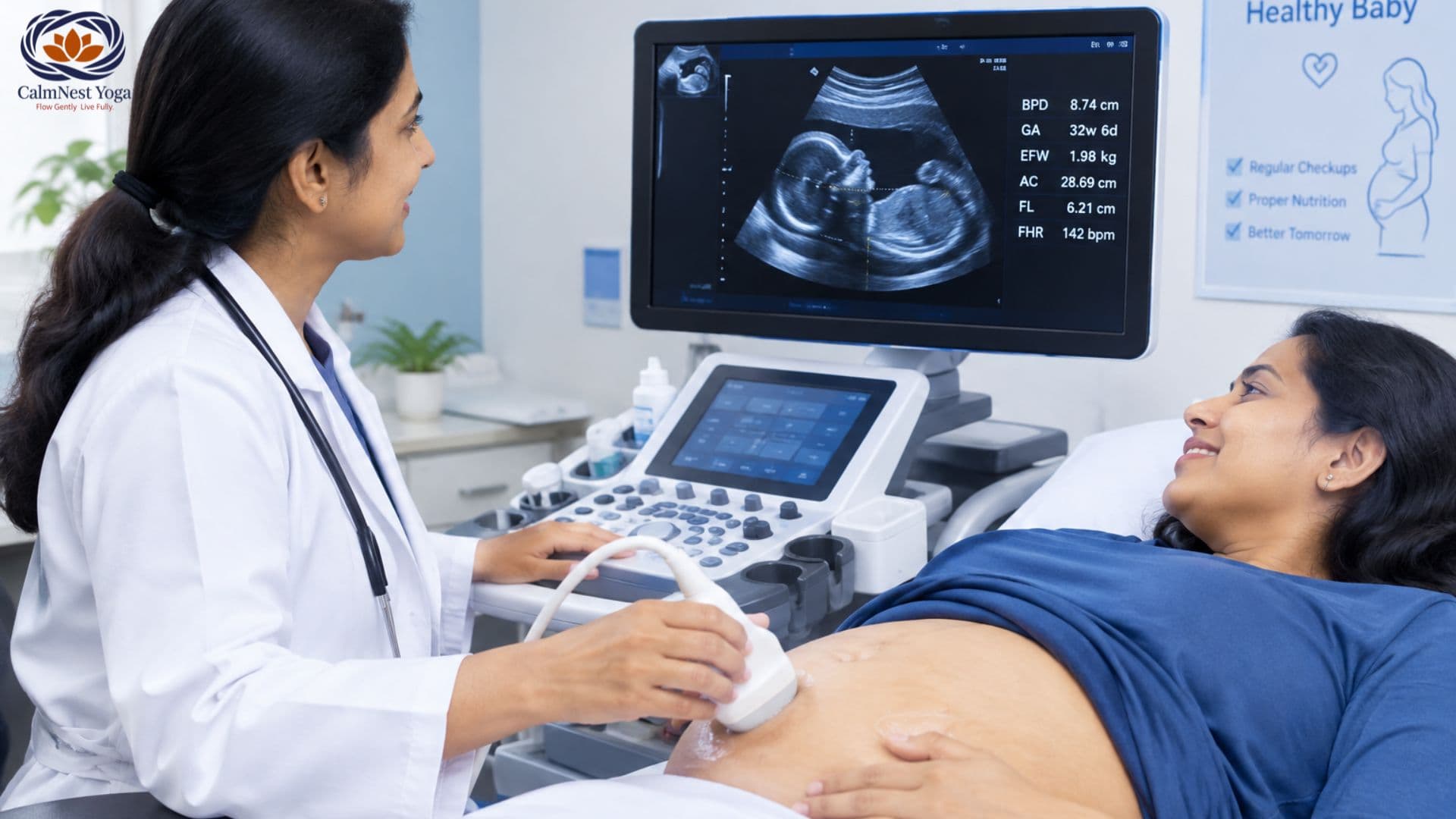
Why do I need a growth scan? When is it done? What measurements are checked? What does a growth scan normal report look like? What do the numbers and percentiles mean? And what happens if the baby is measuring smaller or larger than expected?
Pregnancy involves several important check-ups, blood tests and ultrasound examinations, each designed to monitor a different part of the mother’s and baby’s health. One scan that becomes particularly important later in pregnancy is the growth scan.
If your doctor has recommended a growth scan, you may have questions such as:
Why do I need a growth scan? When is it done? What measurements are checked? What does a growth scan normal report look like? What do the numbers and percentiles mean? And what happens if the baby is measuring smaller or larger than expected?
A growth scan is more than simply estimating the baby's weight. During the examination, the sonographer or doctor may assess fetal measurements, estimated fetal weight, amniotic fluid, fetal position, placenta and, when clinically indicated, blood flow using Doppler ultrasound. Third-trimester ultrasound guidance from the International Society of Ultrasound in Obstetrics and Gynecology (ISUOG) includes assessment of fetal biometry, fetal presentation, placental location, amniotic fluid and relevant Doppler findings.
This guide explains the USG growth scan in detail so that expecting parents can better understand why it is performed and how to read the information in a report.
Important: A growth scan report should always be interpreted in the context of your gestational age, previous scans, medical history and your healthcare provider's assessment. A single number or percentile does not by itself determine whether a pregnancy is healthy.
What Is a Growth Scan in Pregnancy?
A growth scan is an ultrasound examination used to assess the baby's size and growth-related features during pregnancy.
Unlike an early pregnancy scan, which may focus on confirming pregnancy and dating, or an anatomy scan, which primarily examines fetal anatomy, a growth scan is particularly concerned with whether the baby's size is appropriate for the stage of pregnancy and whether other findings provide reassurance about fetal well-being.
During the scan, measurements are taken from different parts of the baby's body. These measurements can then be used to calculate an estimated fetal weight (EFW) and compare fetal size with expected values for that gestational age.
Common fetal biometric measurements include:
- BPD – Biparietal Diameter
- HC – Head Circumference
- AC – Abdominal Circumference
- FL – Femur Length
- EFW – Estimated Fetal Weight
ISUOG guidance identifies BPD, HC, AC and femur diaphysis length among the commonly measured fetal biometric parameters. These measurements can be used in formulas to estimate fetal weight.
However, there is an important distinction:
Fetal size is not exactly the same thing as fetal growth.
A baby's size can be assessed during one ultrasound, but growth is a dynamic process. Comparing measurements over time can provide more information about whether the baby is continuing to grow appropriately.
Why Is a Growth Scan Done?
The main purpose of a growth scan is to assess whether the baby is growing appropriately for the gestational age and to look for findings that may require closer monitoring.
Your doctor may recommend a growth scan for several reasons.
1. To Check the Baby's Growth
The baby's head, abdomen and thigh bone are measured during the ultrasound.
These measurements help estimate the baby's size and weight.
The estimated fetal weight is then compared with a reference range for the baby's gestational age. The result may be expressed as a percentile or Z-score.
For example, a report may mention an estimated fetal weight around the 50th percentile. This means that, according to the particular reference chart being used, the estimated size is around the middle of the reference distribution for that gestational age.
Percentiles should not be interpreted as grades or marks.
2. To Identify Babies That May Be Smaller Than Expected
Some babies are naturally smaller than others.
Being small does not automatically mean that something is wrong.
However, some small babies may have fetal growth restriction (FGR), in which the baby has not reached its expected growth potential.
This distinction is important because a small but otherwise healthy baby and a baby whose growth is restricted may require different approaches to monitoring.
ISUOG notes that fetuses below the 10th percentile are generally categorized as small for gestational age (SGA), but being small does not automatically mean pathological growth restriction.
3. To Check for Excessive Fetal Size
A growth scan can also identify a baby who is estimated to be larger than expected for gestational age.
A larger estimated fetal size can have different explanations, including normal parental or familial variation and certain maternal or pregnancy-related factors.
The ultrasound estimate is not a guaranteed prediction of the baby's birth weight.
4. To Assess Amniotic Fluid
The amount of amniotic fluid around the baby can also be assessed during a growth-focused ultrasound.
Depending on the clinical situation and local protocol, this may involve measuring the deepest vertical pocket (DVP) or amniotic fluid index (AFI).
ISUOG describes DVP and AFI as methods for assessing amniotic fluid volume and notes that the deepest vertical pocket can be used to identify reduced or increased fluid.
Both too little and too much amniotic fluid may need further evaluation depending on the circumstances.
5. To Assess Placental and Fetal Well-being
The scan may also document the location of the placenta and other relevant pregnancy findings.
In some pregnancies, the doctor may recommend Doppler ultrasound to assess blood flow.
Doppler is particularly useful when there is a clinical concern such as suspected fetal growth restriction, hypertension or pre-eclampsia, rather than being automatically necessary for every uncomplicated pregnancy.
6. To Monitor a Previously Identified Concern
A growth scan may be recommended when an earlier examination, maternal condition or previous pregnancy history indicates that additional monitoring would be useful.
Depending on the situation, doctors may follow growth over time rather than relying on one ultrasound.
There is no single week at which every pregnant woman must have a growth scan.
The timing depends on the pregnancy, the reason for the examination and the protocol followed by the treating healthcare team.
Growth-focused ultrasound examinations are commonly performed during the third trimester, but the exact timing may vary.
For example, your doctor may recommend a scan because:
- The baby appears smaller or larger than expected
- There is a concern about fetal growth
- You have high blood pressure or pre-eclampsia
- There is a maternal medical condition that requires closer monitoring
- There has been a previous pregnancy complication
- There are concerns about the placenta or amniotic fluid
- The pregnancy involves twins or multiples
- Previous ultrasound findings require follow-up
- Your doctor wants to monitor growth at intervals
Current ISUOG third-trimester guidance covers assessment of fetal biometry, amniotic fluid, placenta, fetal presentation and Doppler findings where appropriate.
Therefore, instead of focusing only on a particular week, it is better to ask:
“Why has my doctor recommended this scan, and what are they specifically monitoring?”
That question gives you much more useful information than the scan week alone.
What Happens During a USG Growth Scan?
A USG growth scan is generally performed using an ultrasound transducer over the abdomen.
Ultrasound uses sound waves to create images of the baby and surrounding structures.
During the examination, the sonographer may take several measurements.
Depending on the purpose of the examination, the scan may assess:
- Baby's head
- Baby's abdomen
- Femur
- Estimated fetal weight
- Fetal position
- Placental location
- Amniotic fluid
- Fetal heart activity
- Relevant blood flow using Doppler, when indicated
The exact contents of the examination can vary according to the pregnancy and the clinical question.
The scan is generally non-invasive and does not require an injection.
Your healthcare provider may also compare the current findings with earlier ultrasound measurements.
What Measurements Are Checked in a Growth Scan?
Understanding the abbreviations on your report can make the results much easier to follow.
BPD – Biparietal Diameter
BPD measures the width of the baby's head from one side to the other.
It is one of the biometric measurements used in fetal assessment.
HC – Head Circumference
HC measures the circumference of the baby's head.
It provides information about fetal head size and is one of the measurements used in estimating fetal weight.
AC – Abdominal Circumference
AC measures the baby's abdominal circumference.
This is particularly important in fetal growth assessment because changes in abdominal size can provide useful information about fetal growth.
FL – Femur Length
FL measures the length of the baby's thigh bone.
Like the other measurements, it is interpreted in relation to gestational age and the overall fetal assessment.
EFW – Estimated Fetal Weight
The measurements are used in an ultrasound formula to calculate an estimated fetal weight.
It is important to remember the word estimated.
An ultrasound does not place the baby on a weighing scale. Instead, the baby's measurements are entered into a mathematical formula to estimate weight.
ISUOG recommends appropriate fetal biometry and established formulas for estimating fetal weight, with the result interpreted against an appropriate reference standard.
What Does a Growth Scan Normal Report Mean?
Many parents search for a growth scan normal report because they want to understand what a reassuring scan should contain.
A normal or reassuring report generally means that the findings assessed during the examination are considered appropriate for the pregnancy according to the reference standards and clinical context used by the reporting doctor.
A report may include information such as:
- Gestational age
- Fetal presentation
- BPD
- HC
- AC
- FL
- Estimated fetal weight
- EFW percentile
- Placental location
- Amniotic fluid assessment
- Fetal heart activity
- Doppler findings, if performed
- Other observations relevant to the pregnancy
There is no single universal “perfect” growth scan report.
The wording and reference ranges can differ between institutions and clinical situations.
A report saying “growth corresponds to gestational age” is generally reassuring, but your obstetrician should interpret it alongside your previous scans and overall pregnancy history.
Understanding Growth Scan Results and Percentiles
One of the most confusing parts of a report can be the percentile.
You may see statements such as:
EFW – 25th percentile
or
AC – 45th percentile
or
EFW – 75th percentile
What does this mean?
A percentile compares a measurement with measurements from a reference population at the same gestational age.
For example, a measurement around the 50th percentile is approximately in the middle of the reference distribution.
A percentile below the middle is not automatically abnormal.
Similarly, being above the middle does not automatically mean there is a problem.
The 10th percentile is an important clinical threshold used in the assessment of babies who may be small for gestational age, but fetal growth restriction is more nuanced than simply looking at one percentile.
This is why you should avoid interpreting a single number without your doctor.
What Is the Difference Between Small Baby and Fetal Growth Restriction?
This is one of the most important concepts to understand.
A baby may measure small because of normal biological variation.
For example, some healthy babies are naturally smaller because of parental characteristics or other factors.
Another baby may be small because the placenta is not supplying sufficient nutrients and oxygen, or because another pregnancy-related condition is affecting growth.
The second situation may represent fetal growth restriction (FGR) and requires appropriate monitoring.
Therefore:
Small does not always mean unhealthy.
And:
A normal-looking weight estimate does not necessarily provide the complete picture of fetal well-being.
Doctors may consider:
- Estimated fetal weight
- Abdominal circumference
- Growth trajectory
- Amniotic fluid
- Doppler findings
- Maternal blood pressure
- Other maternal conditions
- Previous ultrasound findings
- Clinical symptoms
ISUOG specifically emphasizes that fetal size at a single time point and fetal growth over time are different concepts.
Why Comparing Two Growth Scans Can Be Important
Suppose a baby's estimated weight is within the expected range today.
That is useful information, but it does not necessarily tell the complete story about growth.
A subsequent scan can help determine whether the baby has continued to grow appropriately.
This is why your doctor may compare:
Previous scan → Current scan → Growth pattern
rather than looking at today's number alone.
However, scans should not necessarily be repeated too frequently simply to see whether the baby has grown. The appropriate interval depends on the clinical reason for monitoring.
ISUOG notes that meaningful assessment of interval growth requires measurements separated by an appropriate period; its guidance discusses at least three weeks between measurements when assessing deviations in growth.
Your obstetrician will decide the appropriate timing based on your circumstances.
What Does Doppler Mean in a Growth Scan?
Sometimes a growth scan includes a Doppler study.
Doppler ultrasound evaluates blood flow through specific vessels.
In pregnancies where fetal growth restriction is suspected, an umbilical artery Doppler can provide information about placental circulation and help doctors assess the baby's condition.
ISUOG states that umbilical artery Doppler is particularly used in the management of fetal growth restriction and can help distinguish babies who are small but healthy from those with pathological growth restriction.
Depending on the clinical situation, other Doppler measurements may also be considered.
Importantly, Doppler is not automatically required for every low-risk pregnancy. ISUOG notes that routine umbilical artery Doppler has limited information value in low-risk pregnancies without a specific indication.
So if your report does not contain Doppler measurements, that does not automatically mean something was missed.
What Does Amniotic Fluid Mean on the Report?
Amniotic fluid surrounds the baby and plays several important roles during pregnancy.
A growth ultrasound may describe the fluid as:
- Adequate or normal
- Reduced
- Increased
It may also provide measurements such as:
AFI – Amniotic Fluid Index
or
DVP – Deepest Vertical Pocket
ISUOG guidance describes both approaches and gives reference thresholds for deepest vertical pocket measurements.
If your report mentions reduced or increased fluid, your doctor may recommend additional monitoring depending on the degree of the finding and the overall pregnancy situation.
What Can Cause a Baby to Measure Small?
There are many possible explanations for a baby measuring smaller than expected.
Some babies are simply constitutionally small and healthy.
Other possible factors can include:
- Placental problems
- Maternal hypertension
- Pre-eclampsia
- Certain maternal medical conditions
- Problems affecting placental blood flow
- Multiple pregnancy
- Some fetal conditions
- Incorrect pregnancy dating
This is why doctors do not diagnose fetal growth restriction based only on an isolated estimated weight.
Accurate gestational age is particularly important when interpreting fetal measurements. ISUOG recommends establishing pregnancy dating using reliable early-pregnancy methods when possible and avoiding unnecessary changes to the established due date based on later scans.
What If the Baby Measures Larger Than Expected?
A baby may also measure larger than expected.
Again, an ultrasound estimate does not automatically mean there is a medical problem.
The doctor may consider:
- Maternal health
- Blood glucose levels
- Family or parental characteristics
- Previous pregnancy history
- The baby's growth pattern
- Other ultrasound findings
The estimated fetal weight should be interpreted as an estimate rather than an exact prediction of birth weight.
Can a Growth Scan Predict the Baby's Exact Birth Weight?
No.
This is an important point for expecting parents.
The estimated fetal weight obtained during ultrasound is calculated from fetal measurements. It is not the same as directly weighing the baby.
There can be a difference between estimated fetal weight before birth and the baby's actual weight at delivery.
Therefore, if your report estimates a particular weight, it should not be interpreted as a guaranteed birth weight.
The value of the scan is much broader than predicting the final number on the weighing scale.
Does a Normal Growth Scan Mean Everything Is Perfect?
A reassuring growth scan is good news, but it does not guarantee that every possible pregnancy or fetal problem is absent.
Ultrasound has specific purposes and limitations.
A growth scan mainly provides information about the findings being assessed at that time.
Your overall prenatal care also involves:
- Regular antenatal appointments
- Maternal blood pressure monitoring
- Appropriate blood tests
- Fetal movement assessment
- Screening and diagnostic tests when indicated
- Monitoring maternal symptoms
- Other ultrasound examinations when recommended
A normal growth scan normal report should therefore be understood as one reassuring part of prenatal care rather than a complete guarantee about the remainder of pregnancy.
Growth Scan vs Anomaly Scan: What Is the Difference?
These two scans are sometimes confused.
Anomaly Scan
The anomaly scan, usually performed during the second trimester, primarily focuses on examining fetal anatomy and screening for structural abnormalities.
Growth Scan
A growth scan focuses more strongly on fetal size, growth-related measurements and other features relevant to fetal well-being.
A third-trimester ultrasound can include fetal biometry, fetal presentation, placenta, amniotic fluid and Doppler assessment where appropriate.
The two examinations can therefore answer different clinical questions.
What Should You Look for in Your Growth Scan Report?
When you receive your report, you may want to identify these sections:
1. Gestational Age
Check the gestational age used for interpretation.
2. Fetal Biometry
Look for:
- BPD
- HC
- AC
- FL
3. Estimated Fetal Weight
Look for the EFW and, where provided, the corresponding percentile.
4. Amniotic Fluid
Look for the assessment of fluid volume or AFI/DVP measurement.
5. Placenta
Check the documented placental location and any comments made by the reporting doctor.
6. Fetal Presentation
The report may state whether the baby is cephalic, breech or in another position.
Remember that fetal position can change during pregnancy, particularly before labour.
7. Doppler
If Doppler was clinically indicated, the report may contain blood-flow measurements and interpretation.
8. Impression or Conclusion
This is often the most useful section because it summarizes the radiologist's or fetal medicine specialist's interpretation.
Rather than focusing on one number, read the conclusion together with your obstetrician.
What Questions Should You Ask Your Doctor After a Growth Scan?
If you are unsure about your growth scan results, consider asking:
- Is my baby's growth appropriate for the gestational age?
- What is the estimated fetal weight percentile?
- Is the abdominal circumference within the expected range?
- Is the amniotic fluid normal?
- Is the placenta functioning as expected based on the findings available?
- Was Doppler required in my case?
- Do the current measurements show appropriate interval growth?
- Do I need another growth scan?
- Is there anything I should monitor at home?
- When should I return for my next antenatal appointment?
These questions can help turn a confusing report into a useful conversation with your healthcare provider.
Does Every Pregnant Woman Need Multiple Growth Scans?
Not necessarily.
The number and timing of ultrasound examinations should depend on the individual pregnancy.
Some women may have an uncomplicated pregnancy and require fewer additional scans.
Others may need serial growth monitoring because of risk factors or an ultrasound finding.
For example, additional monitoring may be considered when there is:
- Suspected fetal growth restriction
- Maternal hypertension or pre-eclampsia
- A previous pregnancy affected by growth problems
- Multiple pregnancy
- Certain maternal medical conditions
- Concern about fetal size
- Concern about amniotic fluid
- Abnormal Doppler findings
Your obstetrician is the appropriate person to determine how often monitoring is necessary.
Are Growth Scans Safe?
Ultrasound is widely used during pregnancy and does not involve ionizing radiation.
A medically indicated ultrasound is generally considered a safe component of prenatal care when performed appropriately.
Doppler ultrasound may also be used when clinically indicated. ISUOG provides specific safety guidance regarding Doppler use in pregnancy and recommends appropriate use by trained professionals.
Pregnant women should nevertheless avoid unnecessary scans simply for reassurance unless recommended by their healthcare provider.
Can Pregnancy Yoga Affect the Growth Scan?
Prenatal yoga does not replace medical monitoring or ultrasound examinations.
However, appropriate prenatal yoga can be part of a healthy pregnancy routine for women who have been medically cleared to exercise.
At CalmNest Yoga, prenatal sessions focus on gentle, pregnancy-appropriate movement, breathing, relaxation and preparation for childbirth.
You can learn more about our Prenatal Yoga Classes and explore pregnancy-focused resources on the CalmNest Yoga website.
If your doctor has advised restricted activity, bed rest or other precautions, follow your medical team's instructions rather than beginning or continuing an exercise programme without clearance.
Growth Scan Results: When Should You Seek Medical Advice?
A scan report should be discussed with your obstetrician if it contains terms such as:
- Low estimated fetal weight
- Small for gestational age
- Fetal growth restriction
- Reduced amniotic fluid
- Increased amniotic fluid
- Abnormal Doppler
- Abnormal placental findings
- Significant change from a previous scan
Do not try to diagnose a pregnancy complication from a Google search or from a single percentile.
Some findings require simple follow-up, while others may require closer fetal surveillance.
If you experience symptoms such as vaginal bleeding, leaking fluid, severe abdominal pain, severe headache, visual changes, sudden swelling, or a noticeable reduction in your baby's usual movements, contact your maternity care team promptly rather than waiting for a scheduled scan.
Why Growth Monitoring Matters for Fetal Well-being
The purpose of fetal growth monitoring is not simply to find out how much the baby weighs.
It is about identifying patterns that may help doctors determine whether the pregnancy needs closer observation.
When a growth concern is identified, doctors may use a combination of:
- Repeat ultrasound measurements
- Fetal growth trends
- Amniotic fluid assessment
- Doppler studies
- Maternal blood pressure
- Maternal symptoms
- Fetal movement
- Other fetal surveillance methods
This combined approach is important because no single measurement can provide a complete picture of fetal well-being.
ISUOG guidance emphasizes that fetal biometry, amniotic fluid assessment, Doppler findings and maternal clinical information can contribute to identifying pregnancies at increased risk.
Final Thoughts: Understanding Your Growth Scan With Confidence
A growth scan can provide valuable information about how your baby is growing and whether additional monitoring may be needed during pregnancy.
Understanding terms such as BPD, HC, AC, FL, EFW, percentile, AFI, DVP and Doppler can make your report much less confusing.
Most importantly, do not judge your pregnancy based on one number.
A growth scan normal report should be interpreted according to your baby's gestational age, the reference chart used, previous ultrasound findings and your overall health.
Similarly, a result that is outside the expected range does not automatically mean that something is seriously wrong. It may simply mean that your doctor wants to monitor the baby more closely.
Your obstetrician or fetal medicine specialist can put the growth scan results into the right clinical context and explain whether the findings are reassuring or require follow-up.
Pregnancy is a journey of regular monitoring rather than a single test. A growth scan is one important part of that journey, helping your healthcare team keep track of fetal growth and make informed decisions when additional care is needed.
Medical Disclaimer
This article is intended for general educational purposes and should not replace advice from your obstetrician, radiologist, fetal medicine specialist or other qualified healthcare professional. Ultrasound findings need to be interpreted in the context of the individual pregnancy, gestational age, previous scans and clinical history.
Sources and further reading
The medical information in this article has been cross-checked against guidance from the International Society of Ultrasound in Obstetrics and Gynecology (ISUOG), including its guidance on fetal biometry and growth, third-trimester obstetric ultrasound and Doppler assessment.
ISUOG – Ultrasound assessment of fetal biometry and growth